Planmed Verity®
The Planmed Verity® Extremity CT Scanner revolutionizes 3D extremity imaging. This compact mobile unit brings 3D imaging to radiology departments, emergency departments, orthopedic clinics and trauma centres for fast diagnoses at the Point-of-Care.
 The Planmed Verity® Extremity Scanner wins the sought-after 2012 Medical Design Excellence Award (MDEA) Gold Winner award, and is also nominated “Best in Show”. MDEA is the premier awards competition for the medical technology community.
![]()
Planmed Verity® Brochure pdf
Planmed Verity® Flyer pdf
CT Arthrography Scientific Paper pdf

Superior image quality serves radiologists, orthopedists, and extremity specialists all alike. With surprisingly low radiation dose of up to 10 times less than that of conventional CT, Planmed Verity® helps to find subtle extremity fractures at the first visit to the clinic.
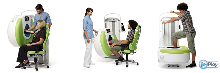
Compact, stand-alone and mobile Planmed Verity® can fit into almost any existing X-ray room and can be easily sited even side by side with other imaging equipment. With adjustable, soft surfaced gantry and dedicated patient positioning trays Planmed Verity® provides versatile patient positioning and optimized patient comfort.
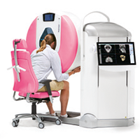
MaxScan™ can be used to image diseases, injuries and defects of:
• Maxillofacial trauma
• Maxillofacial area
• Sinuses
• Mandible
• Temporomandibular joint (TMJ)
• Orbits
• Teeth
• Airways
A non- claustrophobic experience is achieved through convenient positioning and the open design of the gantry.
The effective dose is low because the thyroid gland is not in the primary beam.
Planmed Verity® MaxScan pdf

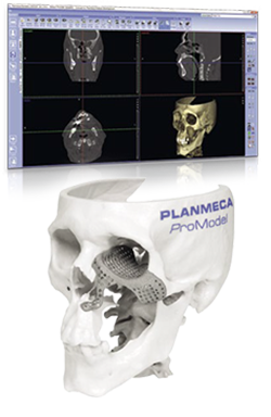
Planmeca ProModel™ offers patient-specific implants, surgical guides and 3D skull models for maxillofacial surgery. Offer you the best tools to succeed in both pre-planning and the surgery itself.
• A unique service for creating patient-specific implants, surgical guides and 3D skull models from CBCT/CT images
• 3D implants are designed in an online session between the surgeon and Planmeca designer
• The implants are designed and manufactured to match any form
• Ordering is quick and easy
• Reduces operation times
• Faster and more precise operations leading to better aesthetic results
• Volumetric imaging with multi-planar reconstruction and volume rendering provide optimal visualization of fractures and deformations
• Superior isotropic resolution of up to 0.2mm (optional 0.1mm high resolution mode)
• Dose up to ten times lower compared to extremity imaging protocols with conventional MDCT (Multi Detector Computed Tomography)
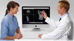
• Revolutionary compact and mobile form for easy positioning in existing X-ray rooms
• Stand-alone system including 3D image acquisition and presentation
• Plugs into a standard GPO electric outlet
• No external cooling requirements, no reinforcement of the floor needed
• Worklist management with touch-screen interface
• DICOM 3.0 conformance including send and export
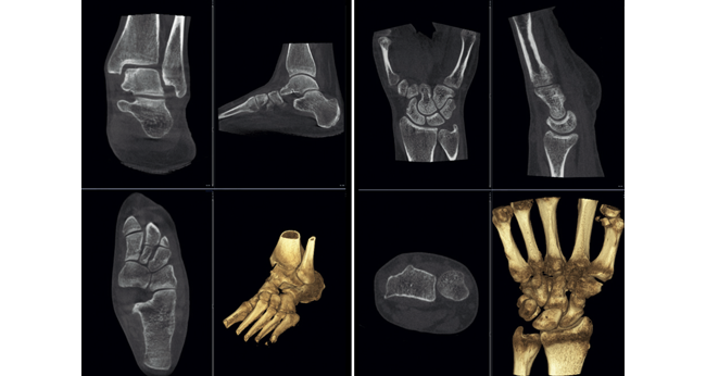
• Easily adjustable, soft surfaced gantry and motorized positioning trays help in finding a comfortable position for various examination procedures
• Easy to access, suits for wheel-chair patients and bed-side imaging
• Compact design prevents claustrophobia and makes the examination more comfortable especially for the elderly and paediatric patients
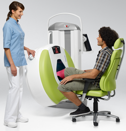
Seated knee, leg, ankle, foot, toes.
Planmed Verity® can always be positioned in a way that is the most convenient for the patient. Fracture healing follow-up is possible without removing the cast.
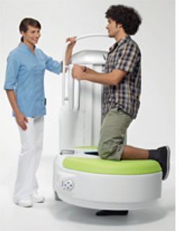
Standing knee, ankle, foot, toes.
3D image of a standing patient shows the anatomy in a natural position, and can reveal problems that are otherwise non-discernible, such as diminished joint space.
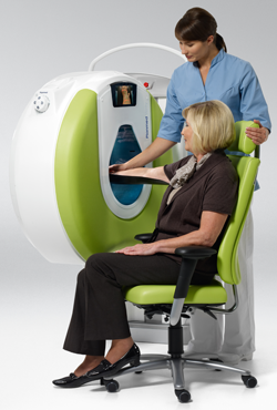
Seated or supine elbow, arm, wrist, hand, fingers.
The versatile positioning also enables imaging directly on a hospital bed. Within the X-ray room, a single person can easily move the unit to the preferred position.
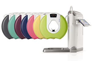 A range of colours are available for selection:
• Dark Blue
• Lilac
• Lime
• Mint
• Sahara Yellow
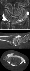
The Planmed Verity Extremity CT scanner was used in a study over a period of 7 months to evaluate arthrography of the wrist. 52 subjects with suspected wrist ligament tears underwent arthrography examinations on the Planmed Verity before normally scheduled MRI arthrography.
The Planmed Verity proved to be very successful in this study providing high spatial resolution at low dose with the potential to provide imaging for orthopedic problems, especially when osteosynthetic material has been used and when an MRI is not available.

