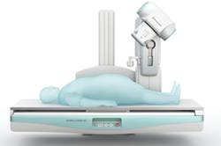
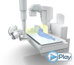
An all in one Remote Controlled Fluoroscopy system providing advanced imaging technology including Tomosynthesis, Slot and RSM-DSA.
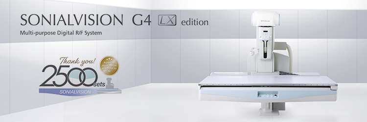
With the Sonialvision G4, Shimadzu offers a total system – one that is useful in a variety of examinations and examination environments, providing high image quality, reduced exposure levels and easy operation.
The Sonialvision G4 is built on Shimadzu”s extensive experience in X-ray imaging and as such offers excellent flexibility and some unique features including:
- • Large Field of View (43x43cm)
- • Patient weight up to 318kg
- • Isocentric tilting
- • Automatic Positioning
- • Urology collimation
- • Offset collimation
- • Virtual collimation
- • Dose reduction technology
- • Image processing engine
- • Fluoroscopy
- • DR radiography
- • Slot radiography
- • Tomosynthesis
- • DSA & RSM DSA
- • DICOM – Worklist, Print, Store, Archive, MPPS
These features make this system ideal for a wide verity of examinations ranging from paediatric to bariatric, DR and series imaging all on one flexible platform.
The large field of view and high image quality is perfect for everything from routine checkups to specialized examinations and observations including gastrointestinal series and video fluoroscopic examinations for swallowing (VF).
The ultra small pixel pitch and high contrast that maximizes the Flat Panel Detector”s performance results in consistent high image quality.
Clear fluoroscopy and radiographic images with enhanced region of interest, suppressed halation near the skins surface and reduced shadows where organs overlap is provided by Shimadzu”s SUREengine, a state-of-the-art multi frequency image processing technology.
SUREengine FAST - The new image processing technology for ERCP and other fluoro based procedures.
SUREengine FAST uses high-speed computational processing to reduce image noise and lag, even at low dose rates. It significantly reduced X-ray dose while maintaining excellent image quality and real time performance required for endoscopic examinations.
Equipped with various functions to effectively reduce exposure levels, the system provides peace of mind for both patients and attending personnel during examinations. This is of particular importance in paediatric and gynecological examinations.
- • Pulsed fluoroscopy
- • Fluoro loop store
- • Grid controlled fluoroscopy
- • Beam hardening filters
- • Asymmetric collimation
- • Virtual collimation
- • Removable grid
- • DAP meter integration
- • High performance Flat Panel Detector
- • Advanced digital imaging
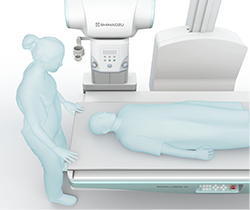
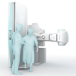 The area around the table is uncluttered and the monitors and local consoles can be freely configured.
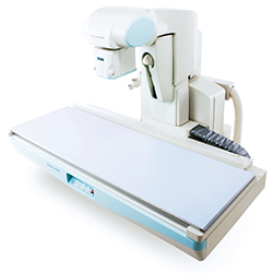
A wide flat table top with high weight capacity ensures patient safety and easy cleaning while full patient coverage is achievable without moving the patient.
With the lowest minimum table height in its class, even frail patients can get on and off easily.
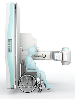
Simply pressing one button will position the system for optimal patient transfer, lowering the table and moving the X-ray tube unit out of the way for patient safety.
The comprehensive tableside controls allow the operator to focus on patient care throughout procedures.
Isocentric tilting means that the table can be tilted without changing the observation position while the large FOV Flat Panel Detector can be positioned close to the end of the table, allowing optimal patient positioning for urological procedures using optical endoscope.
The remote control foot switch adds an ideal user interface providing hands free intuitive operation.
In addition to the standard dose reduction features, utilizing the off-center urological collimation and removing the grid when appropriate further reduce patient dose.
The wide, clean, seamless tabletop offers the highest weight capacity in its class, sufficient for even very heavy patients.
In addition, with the high power X-ray generator, tube and advanced digital image processing bariatric procedures are routinely supported.
 The flexibility of the Sonialvision G4 is further evident in the wide range of acquisition modes available:
- • Pulsed Fluoroscopy
- • DR Radiography
- • Series Acquisition
- • DSA & RSM DSA
- • Video Swallow
Additional advanced technologies ensure that current and future imaging needs are realized:
- • Slot radiography
- • Tomosynthesis
- • DSA & RSM DSA
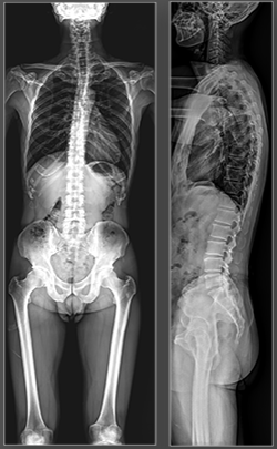
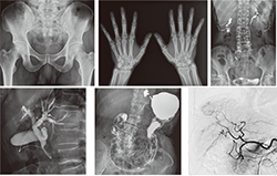
Slot radiography provides a quick and efficient way to acquire long leg and full spine images. Images can be acquired with the table in any position from supine to erect making weigh bearing procedures easy.
The large Flat Panel Detector and the slit beam exposure method results in better patient coverage than traditionally achievable with CR cassettes. In addition, the slot technique produces low dose images with minimal geometric distortion and accurate measurements.
Tomosynthesis is an automated acquisition process that acquires several images while the tube is moving in an arc across the region of interest, similar to the more conventional topographic movement.
The series of images are then processed in a "Cone Beam" or CT-type back projector resulting in a volume of data. The volume of data is then available for investigation by scrolling through the volume in a series of slices.
Slice thickness can be post processed in a similar manor to that used in CT imaging. Slices of interest can be extracted as required for further investigation, printing and DICOM storage.
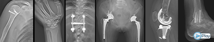
Tomosynthesis provides numerous key advantages including:
- • Low dose
- • Weight bearing
- • Low metal artifacts
- • High spatial resolution
- • High quality imaging in plaster fixation
- • Easy positioning
- • Fast acquisition
- • Large FOV
T-Smart
T-Smart is an advanced version of Tomosynthesis, using Iterative Reconstruction (IR). This results in superior image quality, clearly displaying trabecular detail and less metal artifacts.T-Smart ideal for assessing:
- • Bone/Implant interface assessment
- • Hardware screw loosening
- • Fracture healing status adjacent to fixation plates
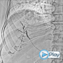
The large field of view means that DSA can be used for examinations of the hepatic artery to the entire lower extremities.
The RSM-DSA filter provides continuous automatic masking resulting in a DSA-like image even when the patient or the table is moved. It produces excellent results where patient movement is inevitable, such as bolus chasing and abdominal examinations, especially when the patient finds it difficult to hold their breath.
 An optional overhead X-ray tube and a wireless Flat Panel Detector can be used in conjunction to provide lateral shoot through and for other out of buck procedures.
In addition, a bucky wall stand with a fixed or wireless Flat Panel Detector can be included to further improve your workflow.
Full DICOM 3 network compatibility ensures patient information can be registered, printed and sent smoothly to your servers.
This provides for an optimised workflow:
- • Register the patient using "Worklist"
- • Select the desired examination (APR)
- • Perform the examination using the pre-programmed examination sets
- • Print DICOM images if desired
- • Send DICOM images to PACS
- • Procedure complete
- • MPPS support throughout the procedure
- • RDSR Radiation dose information report
![]() Benefits of Tomosynthesis for Diagnostic Imaging of Fresh Vertebral Fractures (PDF 1.76MB)
Benefits of Tomosynthesis for Diagnostic Imaging of Fresh Vertebral Fractures (PDF 1.76MB)
Department of Orthopaedic Surgery, Nara City Hospital, Department of Radiology, Division of Medical Technology, Nara City Hospital, Department of Orthopaedic Surgery, Nara Medical University Read more......
![]() Reliability of Diagnosis of Acetabular Dysplasia with Tomosynthesis (PDF 4.45MB)
Reliability of Diagnosis of Acetabular Dysplasia with Tomosynthesis (PDF 4.45MB)
Hironori Ochi, Tomonori Baba, Hiroki Tanabe, Sammy Banno, Yu Ozaki, Yasuhiro Homma, Taiji Watari, Mikio Matsumoto, Hideo Kobayashi and Kazuo Kaneko Department of Orthopaedic Surgery, Juntendo University Read more......
![]() Using Tomosynthesis images for measuring total spine alignment
Using Tomosynthesis images for measuring total spine alignment
Measuring total spine alignment on the Sonialvision system using a combination of Slot and Tomosynthesis imaging. Read more......
![]() Use of Tomosynthesis for Total Hip Arthroplasty (THA)
Use of Tomosynthesis for Total Hip Arthroplasty (THA)
Follow up observations using Tomosynthesis in THA imaging at the Iida Hospital allow assessment of bone fixation. Read more......
![]() Applications of Tomosynthesis in Spine Surgery
Applications of Tomosynthesis in Spine Surgery
Tomosynthesis has become more useful than traditional Tomography in a variety of different regions of interest. Read more......
![]() Can T-Smart Tomosynthesis improve accuracy on THA stability?
Can T-Smart Tomosynthesis improve accuracy on THA stability?
T-Smart Tomosynthesis has greatly improved the quality of THA imaging by reducing the peri-implant artifacts. Read more......
![]() Tomosynthesis for evaluating and treating painful shoulders
Tomosynthesis for evaluating and treating painful shoulders
By creating multi-slice high-resolution images, was beneficial for evaluating the shoulder joint. Read more......
![]() Tomosynthesis in Respiratory Medicine
Tomosynthesis in Respiratory Medicine
A review of Tomosynthesis, CT and radiography in respiratory medicine at the National Cancer Hospital Takehiro Izumo. Read more......
![]() User experience in Cochlear Implant Tomosynthesis
User experience in Cochlear Implant Tomosynthesis
Tomosynthesis clearly shows the relationship between the electrodes and the cochlea senses fro each frequency band. Read more......
![]() Experience in Cochlear Implant Tomosynthesis
Experience in Cochlear Implant Tomosynthesis
Low metal artefacts in Tomosynthesis compared to CT and MRI imaging improve the visualization of the cochlear electrodes. Read more......
![]() Gastrointestinal Tract using Patency Capsules
Gastrointestinal Tract using Patency Capsules
The use of Tomosynthesis to accurately locate and track the Patency Capsule within the abdominal cavity. Read more......
![]() Tomosynthesis changes the evaluation of PLIF bone union
Tomosynthesis changes the evaluation of PLIF bone union
The low metal artefacts in Tomosynthesis imaging from pedicle screws and metal markers make it easier to evaluate the images. Read more......
![]() Experience Using Tomosynthesis (T-smart) at Nara City Hospital
Experience Using Tomosynthesis (T-smart) at Nara City Hospital
T-smart is a new reconstruction algorithm for Tomosynthesis, which reduces the metal artifacts while producing high spatial resolution images. Read more......
![]() The use of Tomosynthesis at Aizawa Hospital
The use of Tomosynthesis at Aizawa Hospital
Because fluoroscopy can be used for accurate positioning, optimal imaging can be obtained for diagnosis. Read more......
![]() The use of Tomosynthesis at Dokkyo Medical University
The use of Tomosynthesis at Dokkyo Medical University
The use of Tomosynthesis for the observation of bone formation after fixation in trauma patients. Read more......
![]() Orthopaedic Surgery Tomosynthesis Sumitomo University Hospital
Orthopaedic Surgery Tomosynthesis Sumitomo University Hospital
Tomosynthesis is extremely useful for diagnostic imaging in orthopedic surgery since it is possible in the erect, sitting or recumbent position. Read more......
![]() Lung Cancer Potential for Low Dose Tomosynthesis
Lung Cancer Potential for Low Dose Tomosynthesis
An investigation into the application of Tomosynthesis in lung cancer screening with a focus on dose reduction. Read more......
![]() Lung Cancer Possibilities Using Tomosynthesis
Lung Cancer Possibilities Using Tomosynthesis
Low dose Tomosynthesis is a useful tool in lung cancer screening. Read more......
![]() The Clinical Utility of Tomosynthesis in Lung Cancer Diagnosis
The Clinical Utility of Tomosynthesis in Lung Cancer Diagnosis
Tomosynthesis offers higher detection capacity for nodules in the lung field compared to general chest radiography. Read more......
![]() Efforts to Reduce Exposure Dose in Chest Tomosynthesis
Efforts to Reduce Exposure Dose in Chest Tomosynthesis
Tomosynthesis is a simple procedure that significantly enhances lesion visibility when compared to conventional chest X-ray imaging. Read more......
![]() Applications of Tomosynthesis to Bronchoscopic Examinations
Applications of Tomosynthesis to Bronchoscopic Examinations
Tomosynthesis simplifies position confirmation for trans bronchial tumor biopsies and permits observations around the peripheral bronchial tubes. Read more......
![]() Usefulness of Tomosynthesis for Orthopedics
Usefulness of Tomosynthesis for Orthopedics
Tomosynthesis enables clear observation of bone fractures, callus formation, fusing, synostosis status and bone and joint structures. Read more......
![]() Clinical Applications to Orthopedics using Tomosynthesis
Clinical Applications to Orthopedics using Tomosynthesis
Tomosynthesis enables clear observation of bone tumors and bone structure that are difficult to observe in plain X-ray examinations. Read more......
![]() Applications of Tomosynthesis for Colon X-ray Examinations
Applications of Tomosynthesis for Colon X-ray Examinations
Tomosynthesis is extremely effective for colon X-ray examinations, especially in areas with complex topographies and lesions. Read more......
![]() Tomosynthesis nearly equals MRI for rheumatoid arthritis
Tomosynthesis nearly equals MRI for rheumatoid arthritis
Tomosynthesis is nearly as good as MRI for detecting signs of rheumatoid arthritis, according to a new study in the February issue of the American Journal of Roentgenology. Read more......
![]() Tomosynthesis and Slot Advanced Imaging
Tomosynthesis and Slot Advanced Imaging
Shimadzu provides advanced imaging on the Sonialvision G4, including Tomosynthesis and Slot radiography. Read more......
![]() Cutting Edge of ERCP - Experience Using the SONIALVISION G4 and Reducing Scattered Radiation Dose Levels (PDF 5.72MB)
Cutting Edge of ERCP - Experience Using the SONIALVISION G4 and Reducing Scattered Radiation Dose Levels (PDF 5.72MB)
Yoshitaka Nakai Department of Gastroenterology, Digestive Disease Center, Kyoto Katsura Hospital Read more......
![]() A Study of Paediatrics Fluoroscopic Examinations (A Method of Dose Reduction with a Removable Anti-Scatter Grid and Selectable Multi Beam Hardening Filter) (PDF 3.26MB)
A Study of Paediatrics Fluoroscopic Examinations (A Method of Dose Reduction with a Removable Anti-Scatter Grid and Selectable Multi Beam Hardening Filter) (PDF 3.26MB)
Yuki Tsuchiya Department of Radiological Technology, Division of Medical Technology, Fussa Hospital Read more......
![]() Low Dose Mode of SUREengine FAST Highly Rated for Use in Biliopancreatic Endoscopy (PDF 1.91MB)
Low Dose Mode of SUREengine FAST Highly Rated for Use in Biliopancreatic Endoscopy (PDF 1.91MB)
Yoshinao Mori, R.T., Yoshikazu Ishii, R.T. Department of Radiology, Kyoto Katsura Hospital (Kyoto Prefecture) Read more......
![]() SONIALVISION G4 High-Performance R/F System and its Tomosynthesis, SLOT Advance, and Bone Density Measurement Applications for High Quality Diagnosis (PDF 1.67MB)
SONIALVISION G4 High-Performance R/F System and its Tomosynthesis, SLOT Advance, and Bone Density Measurement Applications for High Quality Diagnosis (PDF 1.67MB)
Kazuya Takeuchi Takeuchi Rheumatism Orthopedic Surgery Clinic Read more......
![]() SONIALVISION G4 Development of New SUREengine FAST Fluoroscopic Image Processing Technology for Gastroenterological Endoscopy (PDF 1.42MB)
SONIALVISION G4 Development of New SUREengine FAST Fluoroscopic Image Processing Technology for Gastroenterological Endoscopy (PDF 1.42MB)
Tasuku Saito Medical Systems Division, Shimadzu Corporation Read more......
![]() Experience Using the Sonialvision G4
Experience Using the Sonialvision G4
An RF system is one of the systems for which many demands are made due to the verity of intended uses. Improvement in fluoroscopic image quality, as well as shortened examination times, have been essential in improving the quality of medical care at the Koukan Clinic in Kawasaki. Read more......