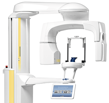
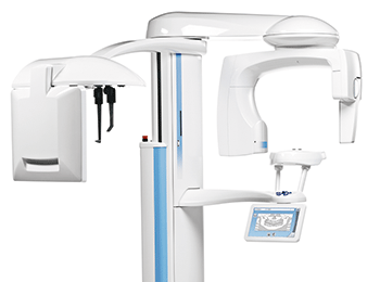
The new member in our Planmeca 3D family
The 3D Plus is the most affordable ‘Radiology class” 3D unit with a very useful 14 cm wide FOV, suitable for your Orthodontic Referrers, with 14 acquisition sizes to choose from.
Whilst not the largest FOV available, this model covers all the standard FOV sizes from Ø40x50 mm on up to Ø200x100 mm for comprehensive implant workup, TMJ studies etc.
In common with the other members of the Planmeca 3D family, 2D Pan & Ceph imaging is integrated into the system.
 Planmeca is the first company in the maxillofacial imaging industry to introduce motion artefact correction, Planmeca CALM™ (Correction Algorithm for Latent Movement), to end users.
- • Never miss a shot
- • Patient movement correction
- • Capture artefact-free images
- • Cancel the effects of patient movement
- • Assure stability in imaging
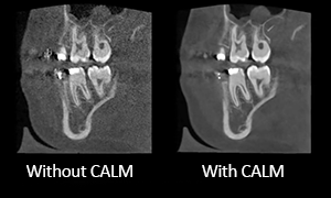 With this new algorithm, patient movement during CBCT acquisition is no longer a reason for retakes. Planmeca CALM™ will not only save time for clinicians but also guard patients from unnecessary exposures.
Upgrade to CALMTM
Planmeca CALM is now available as an update for all Planmeca ProMax® 3D units.Contact us for details about upgrading!
 Ultra Low DoseTM imaging protocols, enable high quality CBCT imaging with an even lower effective patient dose than standard 2D panoramic imaging. This pioneering imaging protocol is based on intelligent 3D algorithms developed by Planmeca and offers a vast amount of detailed anatomical information at a very low patient dose.
This intelligent protocol can be used with all voxel sizes and in all imaging modes from Normal to Endodontic. Using the Planmeca Ultra Low Dose protocol reduces the eff ective patient dose by 75–80%.
![]()
Planmeca Ultra Low Dose
Scientific studies on effective dose.
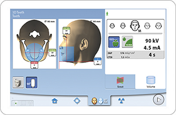
- • Clear and straightforward graphical user interface guides you smoothly through the work process
- • Pre-programmed sites and exposure values for different image types and targets save you time and allow you to focus on your patients
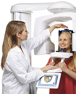
The unique ´Planmeca approach"
- • Effortless positioning with open-face architecture
- • Unrestricted view of your patient
- • No claustrophobic feeling for your patient
- • Fine adjustment using positioning lasers and joystick
- • Verify correct positioning with a scout image
- • Easy wheelchair & Casualty Trolley access
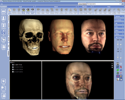
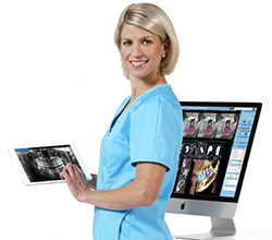 Planmeca Romexis® is an advanced, easy-to-use software suite providing a rich set of tools to meet the imaging requirements demanded by any referrer.
It supports the most versatile range of 2D and 3D imaging modalities & DICOM standard compliance guarantees that images can be processed with 3rd party software or shared via hospital PACS.
Romexis: Read more.
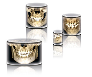 Planmeca ProMax® 3D Plus offers the user a wide variety of volume sizes for applications requiring 3D imaging.
- • Volumes sizes: Ø40x50 mm to Ø200x100 mm
- • A true all-in-one unit: CBCT, 3D photo, 3D model scanning, panoramic and cephalometric imaging, true extraoral bitewings
This wide selection allows optimising the imaging area according to a specific diagnostic task, always complying with the best practices of dentistry and the ALARA (as low as reasonably achievable) principle to minimise radiation.
- • Imaging protocols designed for specific diagnostic tasks, areas, or target sizes
- • Appropriate volume size, resolution, and exposure values
- • Automatic selection and adjustment of the target position
- • Reduced volume sizes for child patients to prevent unnecessary radiation
The new unit also offers a unique combination of three types of 3D data.
The user can acquire a patient CBCT image, 3D face photo and 3D model scan (impression or plaster cast scan), which are combined in one software suite. A virtual patient is created for different clinical needs.
Planmeca is the first to introduce this type of a concept, available for all Planmeca”s 3D units, and these additional unique features can be offered to your Referrer Dental Practices.
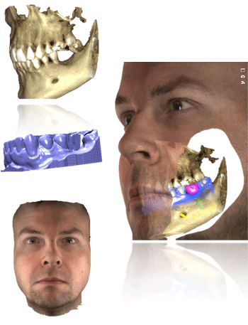
Planmeca ProMax® 3D Family is the first unit to combine 3 different types of 3D data.
- • CBCT image
- • 3D face photo
- • 3D model
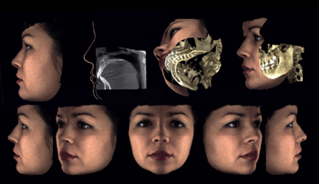 Planmeca's optional ProFace is a unique CBVT imaging upgrade to deliver fully integrated 3D face scanned imaging.
This pioneering integrated system produces a realistic 3D face photo and CBCT image in a single imaging session.
You can also take a separate 3D face photo without exposing your patient to any radiation.
Designed to fulfill the most diverse diagnostic needs of today”s maxillofacial and dental professionals, Planmeca ProFace® is a highly effective tool for pre-operative planning and treatment follow-up.
It”s also ideal for patient motivation and for sharing information with colleagues.
The results can be displayed in 3D or in 2D photo images, & results can be included in PACS files for your Referrers∗
(∗subject to PACS capabilities & configurations)
 Using the information from ProFace, the 3D face photo provides pre and post-operative comparisons. By superimposing images to show deviations with the ability to make measurements and view the relationship between bone and soft tissue. The results can be displayed in 3D or in 2D photo images.
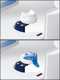 The Planmeca ProMax® 3D family can be used to scan both impressions and plaster casts – an exciting feature that was an industry first for our CBCT units. And with our advanced Planmeca Romexis® software, the digitised models are available immediately and stored for later use.
Scanning a plaster cast to a digital model
Scanning an impression to a digital model
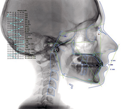
Planmeca Romexis® incorporates a dedicated TMJ imaging module producing detailed images selectable from any angle or projection & with a wide range of selectable slice widths & thicknesses.
The Planmeca Romexis 3D rendering view gives an immediate overview of the anatomy and serves as an excellent patient education tool.
The images can be instantly viewed from different projections or converted into panoramic images and cross sectional slices.
Measuring and annotation tools such as nerve canal tracing assist in safe and accurate planning of treatment.
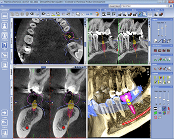 Planmeca Romexis allows easy planning and verification of implant placement using realistic implant models from several manufacturers. Soft tissue surface scan and crown design can be imported and superimposed with 3D X-ray data providing a perfect environment for implant planning.
The virtual treatment plan can be materialised into a real implant guide that can be used to accomplish your treatment exactly as planned.
The dedicated TMJ module provides tools for easy and accurate diagnosis of the TMJ area. The size, location and alignment of the projections can be freely defined and separate views are provided for both TMJs for easy side by side comparison of anatomy.
 Unique ‘low dose” 2D imaging with optimized focus.
A unique SmartPan imaging system uses the same 3D sensor as used for panoramic imaging. This eliminates need to change sensors.
The SmartPan system automatically calculates 10 different panoramic curves in 2 mm shifts from the panoramic exposure data, automatically adjusting the sharpness of one layer.
The user can browse between the panoramic images and select the most suitable for diagnosis after the exposure.
 Using SmartPan you can also select the ‘Pan & TMJ” mode which produces an optimized view of the jaw & the TMJ”s with one low dose acquisition. Both temporomandibular joints will be showing in the image in an optimal and adjustable projection angle.
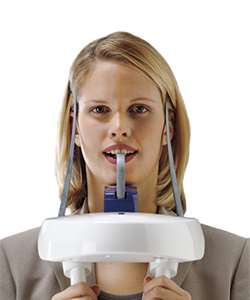
 With Planmeca ProMax Cephalostat imaging is easier and more accurate than ever before. Just move the digital sensor from Pan to Ceph, or select the two fixed digital sensors option.
The functionally designed, easy-to-use head support guarantees accurate patient positioning in all cephalometric projections.
The carbon fibre ear posts and nasal support are extremely durable, hygienic, and fully transparent to radiation.
 ProCeph ‘one shot” DR flat panel cephalostat takes the image with one short exposure, giving excellent ‘motion free” diagnostic image quality with low dose.
- • Available on all Planmeca ProMax, ProMax 3D Classic, ProMax 3D Plus and ProMax Mid models
- • Image size from 20 x 25 to 30 x 25 cm
- • Exposure time 1 s – no motion artifacts
- • High image quality
- • Low dose
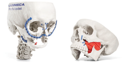 The image data can also be used for ordering Planmeca ProModel, a patient specific physical model that serves as a beneficial tool for preoperative planning of advanced implant, oral and maxillofacial surgeries.
Planmeca uses the precise, free-flowing, computer-controlled SCARA (Selectively Compliant Articulated Robot Arm) arm construction can produce any movement pattern required. This enables accurate and reliable volume positioning and volume diameter adjustment, reducing the amount of radiation your patients are exposed to.
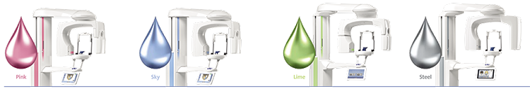
Complement the splendid design of your Planmeca ProMax® 3D X-ray unit by giving it a personal touch with your favorite colours. Select the perfectly matching shades from our exquisite and inspiring collection and create the looks of your dreams!