OPG 3D imaging refined

Planmeca Romexis is comprehensive software for acquiring, viewing and processing 2D and 3D images. All patient images are conveniently processed in a single package software making Planmeca Romexis a powerful tool for studies with multiple image types. Planmeca iRomexis mobile application also enables you to take your work on the road or to quickly share studies with other professionals around the world.
![]()
Planmeca Romexis Brochure
Planmeca Goes Apple

Planmeca Romexis supports different workflows from high patient turnover 2D-imaging to advanced specialist 3D treatment planning. With simplicity as a leading design principle Planmeca Romexis offers easy-to-use tools guaranteeing that the software can be used with minimal training.
Planmeca Romexis offers best in class integration with other systems allowing you to freely utilize 3rd party products at your clinic. TWAIN standard and DICOM standard compliance ensure that Planmeca Romexis can be used effortlessly with most systems.
Full support for both Windows and Apple Macintosh operating systems provides additional freedom in operating your clinic.
 Planmeca Romexis is the software of choice for viewing and processing 2D images acquired with Planmeca”s X-ray units. All patient images can be viewed conveniently in a single user interface minimising clutter on the desktop. The intuitive interface guarantees that the user will feel comfortable using the software from day one. Turn your work into detailed printouts, hand it out with free Planmeca Romexis Viewer software or send it to mobile devices for instant viewing. Fully DICOM compatible software ensures that most features in hospital PACS imaging systems are supported.
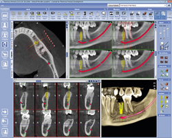
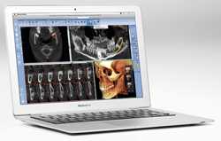
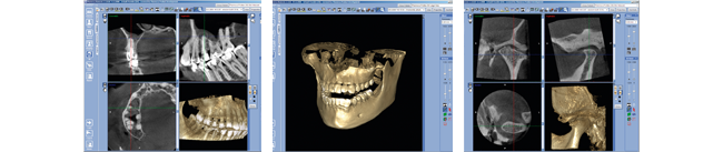 Planmeca Romexis is a comprehensive software solution for acquisition, viewing, and processing of 3D radiographs, 3D photos and intraoral surface scans. The powerful combination of these modalities provides the most accurate information of patient anatomy for different needs. Planmeca Romexis software offers specially designed tools for implantologists, endodontists, periodontists, maxillofacial surgeons and radiologists.
The Planmeca Romexis 3D rendering view gives an immediate overview of the anatomy and serves as an excellent patient education tool. The images can be instantly viewed from different projections or converted into panoramic images and cross sectional slices. Measuring and annotation tools such as nerve canal tracing assist in safe and accurate planning of treatment.
 Planmeca Romexis allows easy planning and verification of implant placement using realistic implant models from several manufacturers. Soft tissue surface scan and crown design can be imported and superimposed with 3D X-ray data providing a perfect environment for implant planning. The virtual treatment plan can be materialised into a real implant guide that can be used to accomplish your treatment exactly as planned.
The dedicated TMJ module provides tools for easy and accurate diagnosis of the TMJ area. The size, location and alignment of the projections can be freely defined and separate views are provided for both TMJs for easy side by side comparison of anatomy.
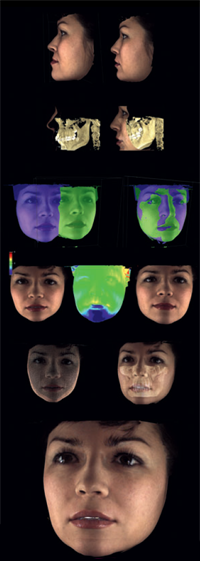
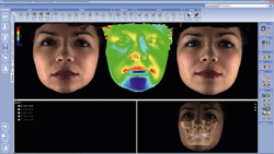 Planmeca Romexis ProFace module allows visualisation of 3D facial photo in relation to facial bone, providing an effective follow-up tool for maxillofacial operations. Careful preoperative planning and thorough study of the facial anatomy facilitates a detailed operation and enhances aesthetic results.
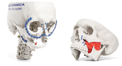 The Planmeca ProModel ordering interface is integrated with the Planmeca Romexis software. Online ordering is made simple, once the desired 3D volume is acquired, all there is to do is to complete the order form in Planmeca Romexis, and the volume is instantly sent to Planmeca”s server.
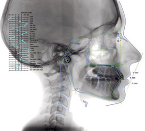 In the Planmeca Romexis® Cephalometric Analysis module, cephalometric analyses and superimpositions can be composed of 2D cephalometric images, facial photos and views of the dental arch.
CEPH analysis is used, for example, in orthodontic growth analysis, diagnosis, treatment planning and monitoring as well as in treatment outcome evaluation. All results can be exported in versatile, customised reports.
- Advanced tools that significantly reduce the time required to carry out a cephalometric analysis.
- Fast and easy analysis using predefined templates, group movement of reference points and automatic calibration.
- Lateral, frontal and arch analyses. The most common 20+ lateral analysis types are included in the software (Ricketts, Bern, McNamara, Downs etc).
- Flexible superimposition of photos and analyses templates.
- Customisable and versatile reports.
Studies can be quickly converted into multi-page printouts or handed out on free Planmeca Romexis Viewer media. Cases can be seamlessly transferred to mobile devices or partner clinics that also use Planmeca Romexis. DICOM standard compliance guarantees that images can be processed with 3rd party software or shared via hospital PACS.
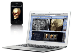 All Planmeca”s X-ray units are fully compatible with Mac OS, including panoramic, intraoral, and 3D CBVT images. Whichever the diagnostic requirement, the images can be acquired, viewed, processed, and stored in a Mac OS environment.
2D and 3D images can be sent to an iPhone and iPad for diagnosis.
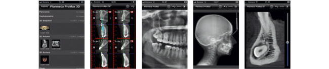
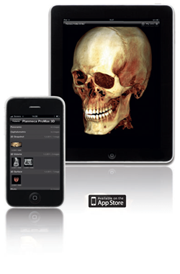 Planmeca iRomexis is a mobile companion application for Planmeca Romexis imaging software designed for Apple iPhone and iPad devices. It allows viewing of 2D and 3D images, 3D renderings and Planmeca ProFace images. Images can be made available for mobile use with Planmeca Online, and downloaded on Wifi and 3G networks wherever you are. Experience a new level of freedom and cooperation with Planmeca iRomexis.
The application is available free at iTunes App Store.
The new service, Planmeca Online, enables sharing of images with mobile devices and between clinics that use Planmeca Romexis. With an account on Planmeca Online, users can send images to any number of supported mobile devices or share images securely with partner clinics that run Planmeca Romexis. Order placement and tracking for Planmeca products are also available. Please visit http://online.planmeca.com for more information.
 Planmeca Romexis Viewer is a light-weight version of Planmeca Romexis desktop software that can be run directly from a CD or DVD without installation.
Every Planmeca Romexis installation can be used to create CDs holding a copy of selected images accompanied with Planmeca Romexis Viewer. Copies of Planmeca Romexis Viewer may be freely distributed. Images are then easily viewed directly from the media or after copying them to the local hard drive for quicker access.
Full support for both Microsoft Windows and Apple Mac OS operating systems.
• Automatically combined CBVT and 3D photo of the patient
• Provides photorealistic documentation
• Multiple tools for measuring, comparing, adjusting, and superimposing images with supreme usability
• Provides an objective foundation for quantifying patient outcomes
• Outstanding for colleague and patient communication
• Images can be exported to 3rd party orthodontic and surgery simulation software
| Pre and postoperative comparisons |
| Measure distances and relations with bone and soft tissue |
| Superimpose images to see the difference |
| Deviation for instant viewing of changes |
| Visualisation of soft tissue in relation to dentin and facial bones |
| Photorealistic documentation |