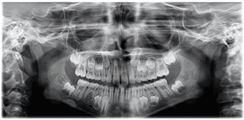
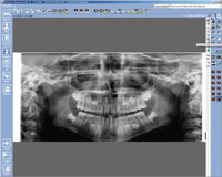
The Planmeca ProMax X-ray unit provides a wide range of extraoral imaging modalities:
• Panoramic imaging for dental arch
• Maxillary sinus imaging
• Temporomandibular joint imaging
• 2D linear tomography (option)
• Cephalometry
• Upgrade option to 3D (11 x 8 cm)

• Versatile presets and imaging modes
• Easy and efficient positioning
• Flexible height adjustments
• Positioning lasers
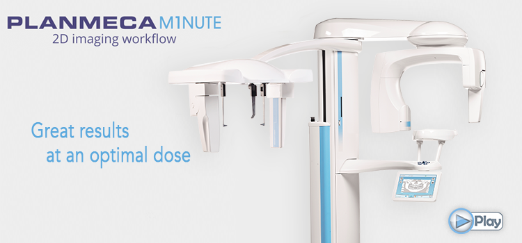
• Great results at an optimal dose
Any Planmeca ProMax film or digital X-ray unit can be easily upgraded to Planmeca ProMax 3D unit by simply changing the imaging sensor and uploading software upgrades.
Both units are also available with 3D face scan.
 Child mode reduces the patient dose remarkably for all programs by reducing the imaging area and exposure values.
In the panoramic program the focal layer can also be narrowed.
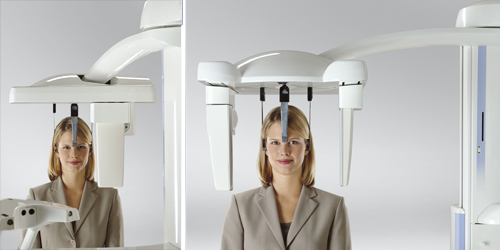
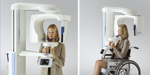 Open patient positioning and side entry minimise errors caused by incorrect patient positioning by allowing you to monitor the patient freely from both the front and side.
Side entry allows easy access for all patients – standing or seated.
If necessary, the patient can even remain seated in a wheelchair or indeed in a casualty trolley.
Patient positioning is assisted by our triple laser beam system, which indicates the correct anatomical positioning points.
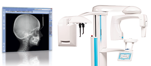
 With Planmeca ProMax Cephalostat cephalometric imaging is easier and more accurate than ever before. By changing the place of the digital sensor the unit switches from panoramic to cephalometric imaging modality. The unit can also be equipped with two fixed digital sensors. The functionally designed, easy-to-use head support ensures easy patient positioning and comfort.
 ProCeph one shot cephalostat takes the image with one single exposure, giving excellent diagnostic image quality with low dose.
- Available on the Planmeca ProMax, ProMax 3D, ProMax 3D s and ProMax Mid models
- Image size from 20 x 25 to 30 x 25 cm
- Exposure time 1 s – no motion artefacts
- High image quality
- Low dose
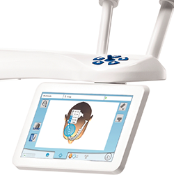 Clear commands and graphic icons make the graphical user interface (GUI) very easy to use which allows for the user to focus on positioning and communicating with the patient. When imaging is completed a preview image shows on the main display.
The unique digital Dynamic Exposure Control (DEC) automatically adjusts the exposure values individually for each patient based on their anatomic structure and bone density. DEC clearly improves the image quality by producing images of more consistent brightness and contrast. DEC is available both for panoramic and cephalometric imaging.
Automatic Exposure Control (AEC) helps to achieve the optimum darkness and contrast. AEC is available both for panoramic and cephalometric imaging.
With the unique Autofocus feature patient positioning is practically error-free. Automatically positioned focal layer uses a brief scout exposure reducing retakes remarkably.
 In addition to the standard panoramic programs, several specialised programs are available.
This includes a child mode where the imaging area and exposure values in all programs are reduced and in the panoramic program focal layer can be narrowed.
This reduces the patient dosage by 35%, while providing full diagnostic information.
Special Image Programs......
Planmeca ProMax tomography programs provide correct topographic information and all required images for the analysis, planning, and follow-up of implant and surgery procedures.
The Planmeca ProMax tomography system produces clear tomographic slices of any part of the maxilla, mandible, or temporomandibular joints. The cross-sectional or longitudinal tomographs can be adjusted to any specific angle and the constant 1.5-fold magnification factor and combination programs enable exact measurements.
Tomography programs......
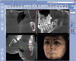 Planmeca's optional ProFace is a unique CBVT imaging upgrade to deliver integrated 3D face scanned imaging.
Designed to fulfil the most diverse diagnostic needs of today”s maxillofacial and dental professionals, it acquires patient”s facial 3D photo in a radiation-free process giving the medical or dental professional opportunity to plan operations and document the follow-up images.
One single scan generates both a 3D photo and a CBVT volume. Alternatively, the 3D photo can be acquired separately in a completely radiation-free process: the lasers scan the facial geometry and the digital cameras capture the colour texture of the face.
Planmeca ProMax utilises unique SCARA technology (Selectively Compliant Articulated Robot Arm).
SCARA is a revolutionary electro-mechanical construction, providing flexible, precise, and complex movements required in rotational maxillofacial radiography.
SCARA technology is combined with real-time computation of dynamic rotation patterns.
These enable optimised radiography of each patient”s anatomy, meeting with virtually any diagnostic requirement in maxillofacial dentistry.
In order to get accurate and clear panoramic radiographs, the form of the X-ray unit focal layer must follow the actual patient anatomy. In Planmeca ProMax, the form of the focal layer follows the scientifically defined shape of human dental arch and jaw resulting in panoramic radiographs with clearly superior clinical quality. The jaw shape and size varies between individuals according to their size, gender, race, and age. Consequently, a single panoramic focal layer form does not suit for all patients. 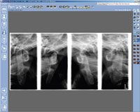
In Planmeca ProMax, the operator may adjust the shape of the focal layer according to the jaw shape and size characteristic to the patient. Planmeca ProMax”s imaging geometry efficiently eliminates redundant shadows and ghost images caused by objects outside the image layer significantly increasing the diagnostic value of panoramic radiographs. The shadow of the cervical vertebrae, which commonly disturbs the clarity of the anterior region, is automatically eliminated by adjusting the amount of radiation in the central incisor region. This computer-controlled correction ensures that there is no loss of image contrast or density.
 The unique design allows an exceptional range of image sizes and formats with field sizes of up to 30 x 27 cm making digital lateral radiographs of the whole skull very easy. With the soft tissue filter applied in the Planmeca Romexis imaging software the images can be viewed with or without the filter.
Romexis: Read more.
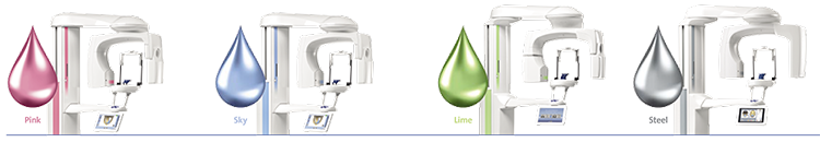
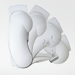
Complement the splendid design of your Planmeca ProMax® 3D X-ray unit by giving it a personal touch with your favorite colours. Select the perfectly matching shades from our exquisite and inspiring collection and create the looks of your dreams!